🏆 Counting down to the European Digital Connectivity Awards 2025!
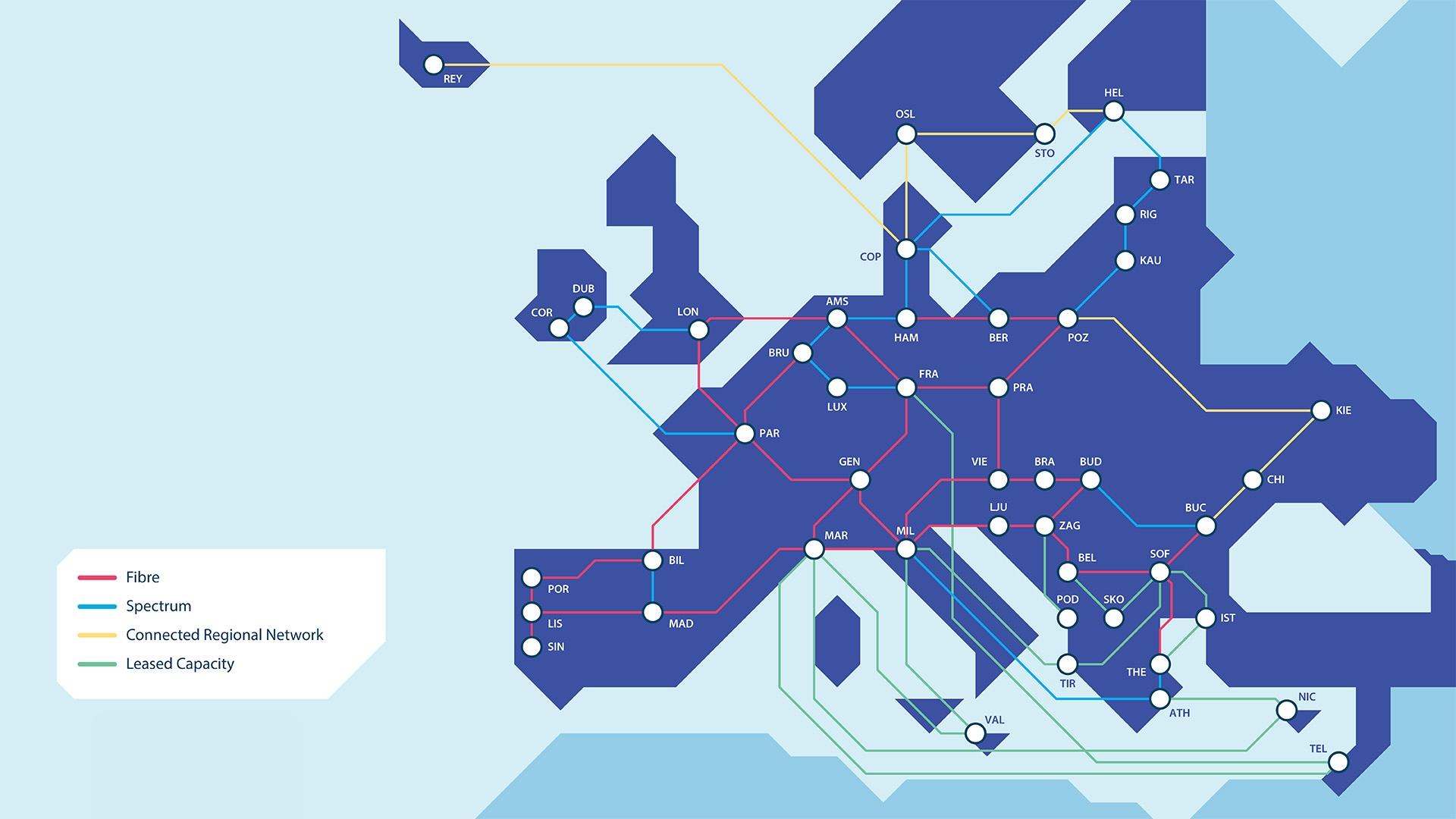
Next week, the winners will be announced—and we’re honoured that GÉANT’s GN4-3N project is a finalist in the “Cross-border and international connectivity” category.
This recognition reflects the collaborative effort of the EU, GÉANT and our community of European NRENs to build a more inclusive, terabit-ready, and fully sovereign network infrastructure for Europe’s #Research & #Education.
🔗 Read more: https://connect.geant.org/2025/07/29/geants-gn4-3n-project-nominated-for-european-digital-connectivity-awards-2025